 |
| Lamprey notochord and vacuolated core cells |
The notochord, a slender elastic-like rod,
is one of four biological features that draw together a wide range of animals
into a single grouping named Chordata. Along with the notochord, the dorsal nerve cord, pharyngeal slits and postanal
tail are characteristics shared by an assemblage that includes both humans
and the sac-like sea squirt. Vertebrates
make up the vast majority of the animals represented but the larval form of the
marine sea squirt gives them admission to this distinguished club, as
well.
 |
| 45 hour old chick embryo with notochord |
The
notochord is a hydrostatic organ with a tough
outer wall enclosing a fluid core. This
gives it lateral elasticity while enabling it to resist any axial
compression. Anchored to this rod, that
extends nearly the length of the organism, is a series of segmented muscles
used, in most instances, to give the animal the means of propulsion through the
water. The contraction of muscles on one
side and then to the next provides alternating lateral pressure against the
surrounding substrate. The resulting
undulating motion propels the animal forward.
Once the muscles relax after contracting on one side of the body the
springy notochord acts to straighten the body out. The notochord acts as the antagonist against
the muscles’ action, enhancing the sweeping of the tail from side to side.
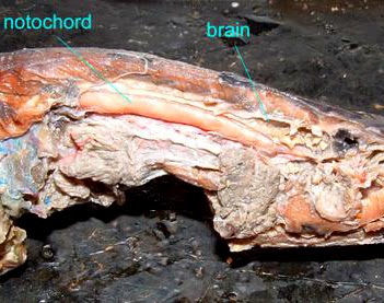 |
| Lamprey notochord extending beneath brain |
The
hydrostatic nature of the notochord prevents the compression of the animal’s
axis which would severely hinder its ability to swim. This pressure is provided by fluid residing
between the notochord’s core cells or by core cells swollen with vacuoles containing fluid. These vacuolated cells are wrapped tight
within a sheath of tough, fibrous tissue.
Under these conditions the inner fluid is held fixed, unable to flow.
 |
| Zebrafish embryo with notochord, segmented muscles |
The
notochord may persist in more primitive chordates but in the case of bony fish
and terrestrial vertebrates this rod is replaced by the vertebral column. In these instances the notochord appears as a
structure used as a scaffold around which the embryonic body can grow. It makes its appearance early when the
mesodermal layers at the dorsal midline differentiates into the chordamesoderm tissue.
This gives rise to the notochord as well as further stimulating the
differentiation of the overlying ectoderm into producing the central nervous
system. It is consequently above the body’s main
central cavity, or coelom, and beneath the
dorsal nerve cord.
 |
| Human vertebrae with notochord derived discs |
The
notochord does not necessarily disappear.
In adult mammals it has transformed into a series of intervertebral
disks. These form circular pads that lie
between the successive vertebrae. Each
pad is a fibrocartilage tissue that encloses a gel-like core, called the nucleus pulposus, providing a cushion between the
connected bony vertebrae. If you’ve ever
suffered a slipped, ruptured or crushed disc you know how important these
structures can be to your general well-being and a healthy frame of mind.

No comments:
Post a Comment